surgery after epidural hematoma and what to exoect
Continuing Education Activity
An epidural hematoma (EDH) is an extra-axial collection of blood within the potential space between the outer layer of the dura mater and the inner table of the skull. It is confined past the lateral sutures (particularly the coronal sutures) where the dura inserts. It is a life-threatening condition, which may require immediate intervention and tin exist associated with significant morbidity and mortality if left untreated. Rapid diagnosis and evacuation are important for a good outcome. This activity examines when this condition should exist considered on differential diagnosis and how to properly evaluate information technology. This action highlights the function of the interprofessional team in caring for patients with this condition.
Objectives:
-
Identify the epidemiology of epidural hematomas.
-
Explain when an epidural hematoma should exist considered on differential diagnosis.
-
Explicate how to manage an epidural hematoma.
-
Explicate the need for a well-integrated, interprofessional team approach to meliorate care for patients with epidural hematomas.
Admission costless multiple option questions on this topic.
Introduction
An epidural hematoma (EDH) is an extra-centric collection of claret within the potential space between the outer layer of the dura mater and the inner table of the skull. It is confined by the lateral sutures (specially the coronal sutures) where the dura inserts. It is a life-threatening status, which may require immediate intervention and tin can be associated with meaning morbidity and bloodshed if left untreated. Rapid diagnosis and evacuation are important for a good issue.[one][2][3]
Etiology
Information technology occurs in approximately 10% of traumatic brain injuries (TBI) requiring hospitalization. Both traumatic and non-traumatic mechanisms can crusade an epidural hematoma.[4][5]
The bulk of cases related to a traumatic mechanism are a result of caput injury due to motor vehicle collisions, concrete assaults, or accidental falls.
Not-traumatic mechanisms include the post-obit:
-
Infection/Abscess
-
Coagulopathy
-
Hemorrhagic Tumors
-
Vascular Malformations
Epidemiology
An epidural hematoma occurs in 2% of all head injuries and up to xv% of all fatal head traumas. Males are more often afflicted than are females. Furthermore, the incidence is higher among adolescents and young adults. The mean age of afflicted patients is 20 to 30 years, and it is rare after 50 to 60 years of age. As an individual's age advances, the dura mater becomes more than adherent to the overlying bone. This decreases the chance that a hematoma tin develop in the space between the attic and dura.[6]
Pathophysiology
Arterial Injury
Most epidural hematomas result from arterial haemorrhage from a branch of the center meningeal artery. The inductive meningeal artery or dural arteriovenous (AV) fistula at the vertex may be involved.[7][8]
Venous Injury
Up to 10% of EDHs are due to venous bleeding following the laceration of a dural venous sinus.
In adults, up to 75% of EDHs occur in the temporal region. Nevertheless, in children, they occur with similar frequency in the temporal, occipital, frontal, and posterior fossa regions.
A skull fracture is present in the bulk of patients with EDH. These hematomas often nowadays below a fracture of the squamous part of the temporal bone.
If this condition occurs inside the spine, this entity is described as a spinal epidural hematoma.
Based on radiographic progression, information technology can exist classified into one of the following
-
Type I: Acute; occurs on twenty-four hours one and associated with a "swirl" of united nations-clotted claret
-
Type Ii: Subacute occurring between days 2 to four and usually solid
-
Type III: Chronic occurring between days 7 to 20; mixed or lucent appearance with contrast enhancement
History and Physical
The typical presentation is an initial loss of consciousness post-obit trauma, a consummate transient recovery ("ofttimes termed every bit a lucid interval"), culminating in a rapid progression of neurological deterioration. This occurs in fourteen% to 21% of patients with an EDH. However, these patients may be unconscious from the beginning or may regain consciousness later on a brief blackout or may accept no loss of consciousness. Therefore, the presentations range from a temporary loss of consciousness to a blackout. Beware that the lucid interval is non pathognomonic for an EDH and may occur in patients who sustain other expanding mass lesions. The classic lucid interval occurs in pure EDHs that are very large and demonstrate a CT scan finding of active bleeding. The presentation of symptoms depends on how quickly the EDH is developing inside the cranial vault. A patient with a small EDH may exist asymptomatic, but this is rare. Likewise, an EDH may also develop in a delayed style.
A posterior fossa EDH is a rare event. This kind of EDH may account for approximately 5% of all posttraumatic intracranial mass lesions. Patients with posterior fossa EDH may remain conscious until late in the evolution of the hematoma, when they may suddenly lose consciousness, become apneic, and die. These lesions oft extend into the supratentorial compartment by stripping the dura over the transverse sinus, resulting in a significant amount of intracranial bleeding.
This enlarging hematoma leads to eventual summit of intracranial force per unit area which may be detected in a clinical setting by observing ipsilateral pupil dilation (secondary to uncal herniation and oculomotor nerve compression), the presence of elevated blood force per unit area, slowed center rate, and irregular breathing. This triad is known as the "Cushing reflex." These findings may indicate the need for immediate intracranial intervention to prevent central nervous system (CNS) depression and death.
Evaluation
Imaging studies such as a computed tomogram (CT) browse contain the mainstay of diagnosis. Laboratory studies such equally INR, partial thromboplastin time (PTT), thromboplastin fourth dimension (PT), and liver part test (LFT) may be obtained to assess for increased haemorrhage risk or underlying coagulopathies.[9]
CT Scan
CT scan is the most mutual imaging modality to appraise for intracranial bleeding. Its popularity is related to its widespread availability in emergency departments. The majority of EDHs are identifiable on a CT scan. The classic presentation is a biconvex or lens-shaped mass on brain CT browse, due to the express ability of claret to expand within the stock-still zipper of the dura to the cranial sutures. EDHs does not cantankerous suture lines.
Generally, radiologists apply a standard formula for estimating the amount of claret present in an EDH. It is as follows:
ABC/2
A: The maximum hemorrhage diameter on the CT slice with the largest surface area of hemorrhage
B: The maximum diameter 90 degrees to A on the same CT piece
C: The number of CT slices with hemorrhage multiplied past the slice thickness in centimeters
At that place are, nonetheless, other CT findings that may need to exist taken into account when evaluating EDH. For example, connected bleeding may exist indicated by areas of depression density, or a "swirl-sign." The latter may be used for prognosis, and frequently indicates the demand for surgical intervention. If the EDH abuts brain tissue that is hemorrhagic or contused, it may appear shallow, and thus, may be overlooked if the CT browse is not carefully examined.
Several factors may lead to a non-diagnostic CT scan. These are every bit follows:
-
A low-density claret collection may event from astringent anemia (thus leading to misinterpretation).
-
Arterial extravasation may be reduced secondary to astringent hypotension.
-
A positive finding on CT requires that enough blood accumulates for visualization. If the CT is obtained as well soon afterward trauma, there may not be sufficient accumulation for appropriate interpretation.
-
If the EDH is secondary to venous bleeding, blood accumulation may exist wearisome. This could potentially result in difficulty with CT interpretation.
Magnetic resonance imaging (MRI)
Brain MRI is more than sensitive than a CT scan, peculiarly when assessing for EDH in the vertex. It should be obtained when there is high clinical suspicion for EDH, accompanying a negative initial head CT scan.
In the situation of a suspected spinal EDH, a spinal MRI is the preferred imaging modality, equally it affords higher resolution versus a spinal CT.
Angiography
When evaluating EDHs located in the vertex, the healthcare professional person should evaluate for the presence of a dural arteriovenous (AV) fistula that may have arisen from the middle meningeal artery. Angiography may be required to evaluate the presence of such a lesion fully.
Handling / Management
EDH is a neurosurgical emergency. It, therefore, requires urgent surgical evacuation to foreclose irreversible neurological injury and death secondary to hematoma expansion and herniation. Neurosurgical consultation should be urgently obtained as it is of import to intervene within 1 to 2 hours of presentation.[10][11][12]
The priority is to stabilize the patient, including the ABCs (airway, breathing, circulation), and these should be addressed urgently.
Surgical intervention is recommended in patients with:
-
Acute EDH
-
Hematoma book greater than 30 ml regardless of Glasgow coma scale score (GCS)
-
GCS less than 9 with pupillary abnormalities like anisocoria
Operative Direction
In patients with acute and symptomatic EDHs, the handling is craniotomy and hematoma evacuation. Based on the bachelor literature, "trephination" (or burr pigsty evacuation) is oftentimes a crucial grade of intervention if more than advanced surgical expertise is unavailable; it may even decrease mortality. Nonetheless, the performance of a craniotomy, if feasible, can provide a more thorough evacuation of the hematoma.
Non-Operative Management
There is a scarcity of literature comparing bourgeois management with surgical intervention in patients with EDH. All the same, a not-surgical approach may be considered in a patient with acute EDH who has balmy symptoms and meets all of the criteria listed below:
-
EDH volume of less than 30 ml
-
Clot diameter of less than 15 mm
-
Midline shift of less than v mm
-
GCS greater than viii and on physical examination, shows no focal neurological symptoms.
If the decision is made to manage acute EDH not-surgically, close observation with repeated neurological examinations and continuous surveillance with encephalon imaging is required, as the take a chance for hematoma expansion and clinical deterioration is present. The recommendation is to obtain a follow-up head CT scan within six to 8 hours following brain injury.
Differential Diagnosis
-
Intracranial abscess
-
Intracranial mass
-
Seizure
-
Transient ischemic attack
Prognosis
In general, patients with pure EDHs have an splendid prognosis of a functional issue after the surgical evacuation, when it is rapidly detected and evacuated. A delay in diagnosis and treatment increases morbidity and mortality.
EDHs caused past arterial bleeding develop rapidly and can be detected quickly. But those due to a dural sinus tear develop more slowly. Thus, clinical manifestations may be delayed, with a resultant delay in recognition and evacuation. Mostly, an EDH volume greater than l cm prior to evacuation results in a worse neurological outcome and consistent mortality.
Factors that may influence the outcome are equally follows:
-
Patient age
-
Time lapsed between injury and treatment
-
Immediate coma or lucid interval
-
Presence of pupillary abnormalities
-
GCS/motor score, on inflow
CT findings (hematoma book, the degree of midline shift, the presence of signs of agile hematoma bleeding, or associated intra-dural lesions)
Postoperative intracranial pressure (ICP)
Several markers that correlate with a poor prognosis of EDH include the following:
-
A low GCS before surgery, or on arrival
-
Aberrant student exam, in particular, un-reactive pupils (unilateral or bilateral)
-
Advanced age
-
The time betwixt neurological symptoms and surgery
-
Elevated ICP in the post-operative period
Certain head CT findings can correlate with a poor prognosis:
-
Hematoma book of greater than 30 to 150 ml
-
A midline shift greater than ten to 12 mm
-
"Swirl sign" indicating an active drain
-
Associated intracranial lesions (such as contusions, intracerebral hemorrhage, subarachnoid hemorrhage, and diffuse brain swelling)
Complications
-
Mass effect: pinch of the encephalon if bleeding is pregnant
-
Herniation
-
Seizures
Consultations
Post-obit services should be immediately consulted as the outcome is time-dependent:
-
Trauma surgery
-
Neurosurgery
Pearls and Other Issues
-
EDH is a neurosurgical emergency
-
Suspect EDH if there is a history of head trauma leading to a period of loss of consciousness.
-
Patients with small EDH may exist asymptomatic
-
Generally, occurs with a skull fracture, but may occur without a bone fracture
-
EDH does not cantankerous suture lines
-
Although lucid interval is commonly described but not pathognomonic and may occur in patients with expanding mass lesions
-
Patients with EDH may be unconscious, may regain consciousness after a cursory loss of consciousness or may accept no loss of consciousness; Patients with small EDH may exist asymptomatic
-
If rapidly detected and evacuated, the functional outcome is excellent
Enhancing Healthcare Team Outcomes
EDH is a relatively common presentation to the emergency department, and if not diagnosed is associated with high mortality. The condition is all-time managed by an interprofessional team that includes the emergency room medico, the trauma squad, radiologist, neurologist, neurosurgeon, intensivist and the ICU nurses. The condition has been associated with mortality rates in excess of 15%. The key prognostic feature is the level of consciousness at the time of presentation. Both bilateral epidural hematomas and posterior fossa epidural hematomas carry a very loftier mortality. The key to treating this status is preventing it in the beginning place. Healthcare workers should brainwash the public on the importance of head safety equipment when playing sports or while working.[13][14]
Review Questions

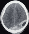
Figure
CT head Epidural Hematoma. Contributed by Scott Dulebohn, Doc

Figure
epidural hematoma afterward trauma. Image courtesy Due south Bhimji MD

Figure
A. A 33-year-erstwhile male involved in a motor vehicle accident with epidural hematomas and skull base fractures presents with right ptosis, proptosis, pulsatile tinnitus, double vision with limitation of motility of right eye in all fields of gaze. Chemosis (more...)

Figure
Calcified EDH. Contributed by Sunil Munakomi, Md
Figure
EDH from sinus origin. Contributed by Sunil Munakomi,MD
References
- one.
-
Rosenthal AA, Solomon RJ, Eyerly-Webb SA, Sanchez R, Lee SK, Kiffin C, Davare DL, Hranjec T, Carrillo EH. Traumatic Epidural Hematoma: Patient Characteristics and Management. Am Surg. 2017 Nov 01;83(11):e438-e440. [PubMed: 30401085]
- 2.
-
Babu JM, Patel SA, Palumbo MA, Daniels AH. Spinal Emergencies in Primary Intendance Practice. Am J Med. 2019 Mar;132(3):300-306. [PubMed: 30291829]
- 3.
-
Kanematsu R, Hanakita J, Takahashi T, Park S, Minami M. Radiologic Features and Clinical Form of Chronic Spinal Epidural Hematoma: Written report of 4 Cases and Literature Review. World Neurosurg. 2018 Dec;120:82-89. [PubMed: 30145384]
- four.
-
Tamburrelli FC, Meluzio MC, Masci M, Perna A, Burrofato A, Proietti L. Etiopathogenesis of Traumatic Spinal Epidural Hematoma. Neurospine. 2018 Mar;xv(1):101-107. [PMC free article: PMC5944636] [PubMed: 29656630]
- v.
-
Fernández-Abinader JA, González-Colón Grand, Feliciano CE, Mosquera-Soler AM. Traumatic Brain Injury Profile of an Elderly Population in Puerto Rico. P R Wellness Sci J. 2017 Dec;36(iv):237-239. [PubMed: 29220069]
- half-dozen.
-
Chicote Álvarez E, González Castro A, Ortiz Lasa M, Jiménez Alfonso A, Escudero Acha P, Rodríguez Borregán JC, Peñasco Martín Y, Dierssen Sotos T. Epidemiology of traumatic brain injury in the elderly over a 25 year menses. Rev Esp Anestesiol Reanim (Engl Ed). 2018 Dec;65(10):546-551. [PubMed: 30054092]
- 7.
-
Burjorjee JE, Rooney R, Jaeger M. Epidural Hematoma Post-obit Cessation of a Direct Oral Anticoagulant: A Instance Study. Reg Anesth Hurting Med. 2018 Apr;43(3):313-316. [PubMed: 29369958]
- 8.
-
Bonow RH, Barber J, Temkin NR, Videtta W, Rondina C, Petroni G, Lujan S, Alanis V, La Fuente G, Lavadenz A, Merida R, Jibaja M, Gonzáles L, Falcao A, Romero R, Dikmen S, Pridgeon J, Chesnut RM., Global Neurotrauma Enquiry Group. The Result of Severe Traumatic Brain Injury in Latin America. World Neurosurg. 2018 Mar;111:e82-e90. [PMC gratis article: PMC5897054] [PubMed: 29229352]
- ix.
-
Flaherty BF, Moore HE, Riva-Cambrin J, Bratton SL. Echo Head CT for Expectant Direction of Traumatic Epidural Hematoma. Pediatrics. 2018 Sep;142(three) [PubMed: 30154118]
- ten.
-
Bhorkar NM, Dhansura TS, Tarawade UB, Mehta SS. Epidural Hematoma: Vigilance beyond Guidelines. Indian J Crit Care Med. 2018 Jul;22(7):555-557. [PMC free article: PMC6069313] [PubMed: 30111936]
- 11.
-
Gutowski P, Meier U, Rohde V, Lemcke J, von der Brelie C. Clinical Outcome of Epidural Hematoma Treated Surgically in the Era of Modern Resuscitation and Trauma Intendance. World Neurosurg. 2018 Oct;118:e166-e174. [PubMed: 29959068]
- 12.
-
Basamh M, Robert A, Lamoureux J, Saluja RS, Marcoux J. Epidural Hematoma Treated Conservatively: When to Expect the Worst. Tin can J Neurol Sci. 2016 Jan;43(one):74-81. [PubMed: 26786639]
- xiii.
-
Aguilar MI, Brott TG. Update in intracerebral hemorrhage. Neurohospitalist. 2011 Jul;ane(three):148-59. [PMC free article: PMC3726132] [PubMed: 23983850]
- 14.
-
Jeong YH, Oh JW, Cho S., Korean Trauma Data Banking company Arrangement Committee. Clinical Effect of Acute Epidural Hematoma in Korea: Preliminary Report of 285 Cases Registered in the Korean Trauma Data Bank System. Korean J Neurotrauma. 2016 Oct;12(2):47-54. [PMC free article: PMC5110918] [PubMed: 27857907]
Source: https://www.ncbi.nlm.nih.gov/books/NBK518982/
0 Response to "surgery after epidural hematoma and what to exoect"
Post a Comment